Revolutionizing Pediatric Cardiology: How Open Source AI and Digital Twins are Transforming Congenital Heart Care at CHOP

The Children’s Hospital of Philadelphia is pioneering a medical revolution by leveraging open-source artificial intelligence to model complex pediatric heart defects in seconds, marking a paradigm shift in how surgeons approach life-saving procedures for children with congenital heart disease. Congenital heart defects (CHD) represent the most common birth defect globally, affecting approximately 1% of all live births. Because every heart presents a unique anatomical fingerprint, the "one-size-fits-all" approach that characterizes many medical devices has historically fallen short for these young patients. By integrating sophisticated AI-driven modeling with GPU-accelerated physics, clinicians at the Children’s Hospital of Philadelphia (CHOP) are now able to conduct "virtual surgeries" that ensure precision, safety, and customized care long before a patient enters the operating room.
The Anatomical Challenge: A Decade of Innovation
The journey from research curiosity to standard clinical practice has been a ten-year evolution. When Dr. Matthew Jolley, a cardiologist and researcher, joined CHOP in 2015, the landscape of pediatric cardiac imaging was vastly different. While 3D echocardiography was emerging, there was a profound lack of specialized tools designed to handle the intricate, small-scale anatomies of children. Standard adult-focused software could not accurately segment the complex defects seen in pediatric patients, leaving surgeons to rely on 2D images and their own spatial intuition.
Dr. Jolley’s team recognized early on that waiting for commercial software vendors to develop niche pediatric tools was not a viable strategy. Instead, they turned to the open-source community. Collaborating with developers, they helped build "SlicerHeart," an extension of the open-source 3D Slicer platform. This enabled researchers to visualize, segment, and analyze 3D medical images with unprecedented clarity. However, the initial process was labor-intensive, often requiring a highly skilled researcher to spend four hours at a workstation to build a single anatomical model. The transition from a research tool to a daily clinical utility required a breakthrough in processing speed, which eventually arrived through machine learning.
The Role of MONAI and AI Segmentation
The integration of MONAI—a medical imaging framework co-founded by NVIDIA—changed the operational capacity of the CHOP team. By utilizing MONAI Label and NVIDIA’s Auto3DSeg implementation, the team trained segmentation networks using historical image-model pairs. The results were transformative: the AI could produce an anatomically precise heart model in seconds, matching the quality of human-segmented models that previously took hours to create.
This leap in efficiency has enabled the modeling service to move from an occasional research project to a routine clinical workflow. CHOP expects to reach approximately 200 modeled cases this year alone. The influence of this approach is now national, with more than 20 children’s hospitals across the United States adopting similar cardiac modeling programs. Boston Children’s Hospital, for instance, has scaled this technology to support more than 50% of its cardiac surgeries, amounting to roughly 500 cases annually.
Physics-Based Simulation: The Next Frontier
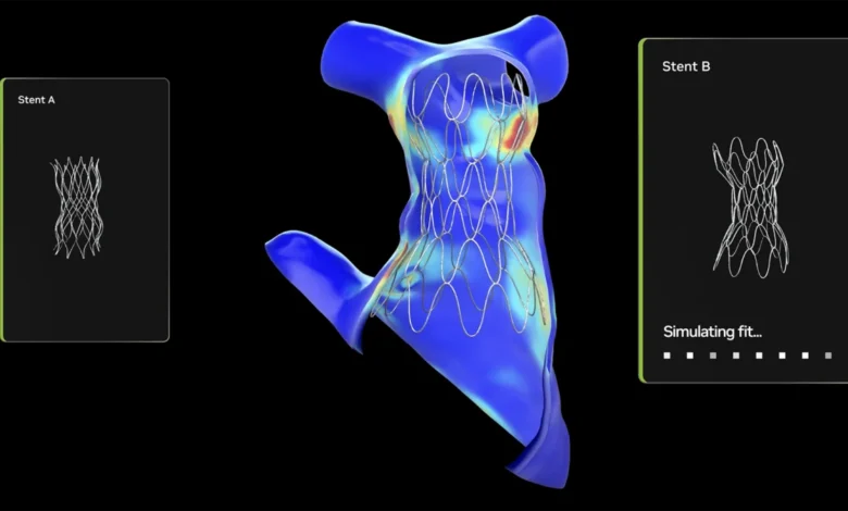
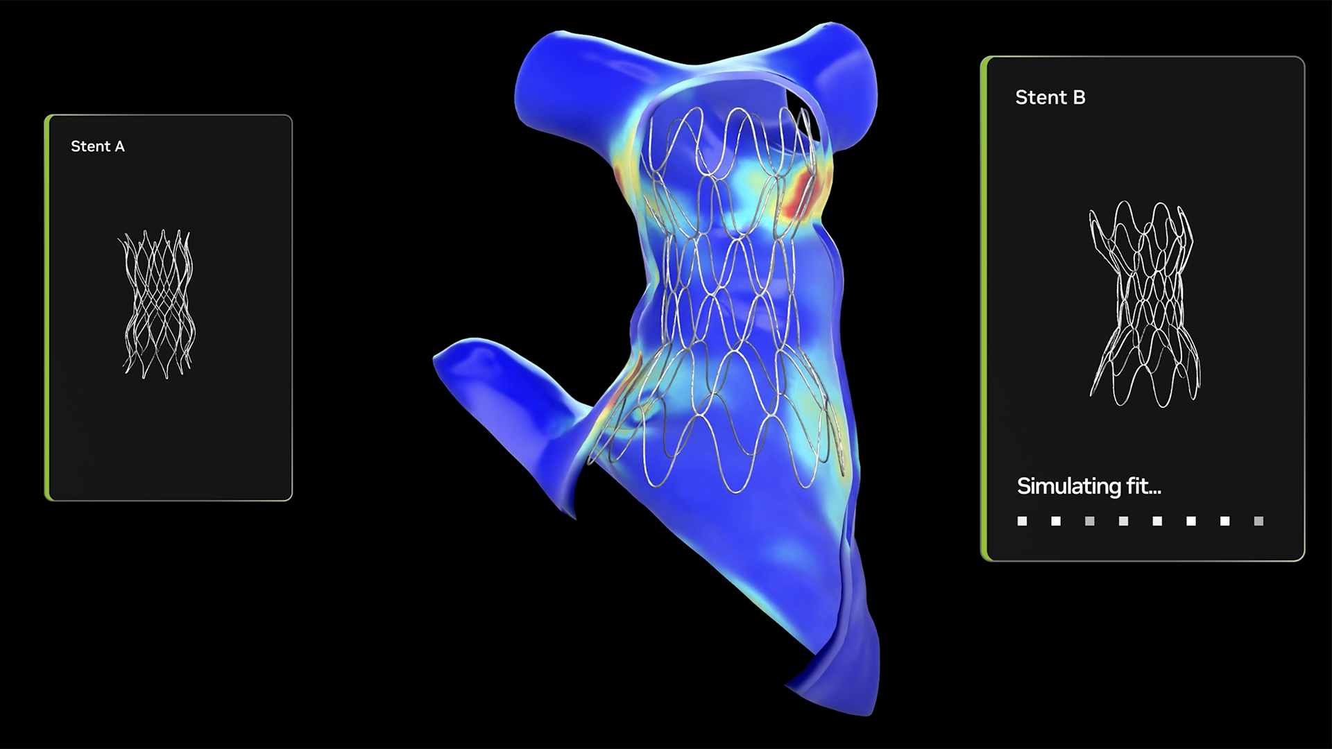
While visual modeling provides a map, it does not always predict the behavior of a device within the heart. To address this, the CHOP team is moving into the realm of predictive biomechanics using the Newton physics engine, built on the NVIDIA Warp Python framework. By running physics simulations on high-performance GPUs, the team can simulate how cardiac tissue will react to specific implants or closure devices.
The implications for surgical planning are significant. In a traditional setting, a surgeon might choose a device based on general sizing guidelines. With the new simulation framework, a clinician can test how two different devices will fit a patient’s specific anatomy, visualizing the stress placed on the cardiac walls in real-time. This reduces the risk of device mismatch—a critical concern in pediatric cardiology where the margin for error is razor-thin. What once required overnight supercomputing time for a single configuration can now be achieved in near-real-time, allowing for same-day clinical decision-making.
Bridging the Gap: OpenUSD and Virtual Reality
Looking toward the future, CHOP is experimenting with NVIDIA Omniverse and OpenUSD to create high-fidelity digital twins of patient hearts. OpenUSD (Universal Scene Description) allows for 3D interoperability, meaning diverse data formats and complex physical solvers can be integrated into a single environment. These digital twins can be exported into virtual reality, where surgeons can "walk through" the anatomy of a patient’s heart.
The integration of vision-language models (VLMs) adds another layer of utility. Surgeons can intuitively query the simulation, asking questions or requesting specific views of the cardiac structure, effectively allowing the AI to act as a clinical assistant. This multi-modal approach ensures that the most complex information—blood flow dynamics, tissue elasticity, and geometric constraints—is presented in a way that is easily digestible for the surgical team.
Economic and Ethical Implications
The adoption of open-source infrastructure is not merely a technical preference; it is an economic necessity. With roughly 2.4 million Americans living with congenital heart disease, the population is large enough to be a significant public health concern but often considered too small and too diverse for traditional, profit-driven medical device companies to justify the R&D costs for highly customized tools.
By relying on open-source frameworks like MONAI and Newton, hospitals can bypass the traditional commercial "bottlenecks." These platforms provide industrial-scale infrastructure that would be impossible for any single hospital to build alone. This collaborative model allows for a "national consortium" approach, where institutions like CHOP, Stanford, and Boston Children’s Hospital can share, build, and refine tools without the friction of proprietary licensing.
A New Standard of Care
The clinical impact of this technology is already being felt. In one notable case at CHOP, a child who had undergone two failed surgeries was saved because a 3D model finally clarified the hidden nature of their ventricular septal defect. The precision afforded by the model allowed the surgeons to identify the exact location of the defect, leading to a successful repair on the third attempt.
As these tools move from the "IDEA Lab" at the Morgan Center for Research and Innovation into standard practice, the definition of surgical preparation is fundamentally changing. The goal is no longer just to "fix" the heart, but to "optimize" the repair for the unique physiology of each child. By democratizing access to high-performance computing and AI, the medical community is proving that, through collaboration, it is possible to provide bespoke, precision care even for the most complex pediatric cases.
The trajectory of this technology suggests that within the next decade, digital twin modeling and real-time biomechanical simulation will become the standard of care in pediatric cardiology. As the open-source community continues to refine these tools, the barriers to high-quality care for children with heart defects are expected to continue to fall, ensuring that "one-of-a-kind" kids receive one-of-a-kind treatment.